MRI – Magnetic Resonance Imaging
Magnetic Resonance Imaging (MRI)
Magnetic Resonance Imaging (MRI) is an imaging modality that is used by physicians for medical diagnosis.
MRI produces images of human anatomy without the use of radiation or x-rays. MRI is a painless procedure that uses strong magnetic fields with radio waves to produce computer-generated images in any plane of the body.
This non-invasive imaging technique can be used as a primary diagnostic tool to visualize the inside of the human body. MRI studies can provide quick and accurate diagnoses for your physician and can reduce the need for further diagnostic or invasive procedures, such as exploratory surgery. MRI is a painless procedure with no side or after effects.
What type of MRI exams are performed?
- Brain
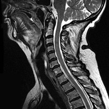
- Spine: cervical, lumbar and thoracic spine
- Musculoskeletal:
- Hip
- Knee
- Ankle
- Foot
- Shoulder
- Elbow
- Hand
- Wrist
- Breast
- Pelvis
- Fibroids, endometriosis, ovaries
- Abdomen
- Liver, pancreas, kidneys, adrenal glands, bowel
How does MRI operate?
MRI images of the body are formed when signals made by body tissues are processed and turned into clinical images. These signals are generated using a safe magnetic field in combination with radio waves of a specific frequency generated by the MRI machine. Different tissue characteristics are translated into different contrast levels on the MRI image.
What to expect during the MRI exam:
The MRI process is painless and takes from 30-60 minutes, depending on the type of study your doctor has requested.
Once you are positioned on the MRI table, the technologist will make sure you are comfortable so it is easy to remain still and breathe normally during the examination.
Each imaging set takes 2-10 minutes, during which time it is necessary to remain as motionless as possible. Once the examination has begun you will hear a knocking sound (which represents changes in the magnetic field). This is a normal part of the imaging process.
Headphones with your choice of music are provided for your relaxation and comfort. You can help optimize your MRI study by relaxing and remaining still during the examination. Some patients may fall asleep during the MRI study.
The MRI table is comfortable and moves inside the center of a large cylinder-shaped magnet, which is open at both ends, ventilated, and well-lighted. There is an intercom so our staff can monitor you and you can communicate with them during the entire scan. At the conclusion of the examination, the technologist will assist you out of the scan room.
What do I need to do to prepare for the MRI exam?
No special preparation is required for the MRI examination. You may eat normally and go about your daily routine. You may continue to take any medications prescribed by your physician, unless otherwise indicated.
Prior to entering the MRI scan room for your examination, you will be asked to leave items that are not compatible with a magnetic field in a secure place outside the scan room. The force of the magnet is strong enough to erase credit cards or disturb some metallic objects in the body.
Some of the items that need to be left outside the room include:
- Coins
- Jewelry
- Watches
- Glasses
- Credit cards
- Hearing aides
- Keys
- Hairpins
- Other metal objects
You may be asked to remove make-up and dentures and to wear a hospital gown to avoid magnetic interference from belt buckles and zippers.
Are you a candidate for MRI?
Check with your physician or the MRI technologist if you have had any brain, ear, eye surgeries, or if you have any of the following:
- Cardiac pacemaker
- Intracranial aneurysm clips
- Cochlear implant
- Neural stimulator (TENS) unit
- Intrauterine device (IUD)
- Implanted drug infusion device
- Foreign metallic objects in the eye
- Shrapnel or bullet wounds
- Permanent eyeliner
If you are pregnant please notify your physician and/or the MRI technologist.
Intravenous contrast
Some MRI studies will require the use of intravenous contrast material to outline vessels and blood flow of the area being evaluated.
If contrast is needed for your examination, a small intravenous line will be placed prior to obtaining the images and the contrast will be infused during the study. A blood test of renal function may be performed before contrast imaging is approved. This may be necessary if there is a history of diabetes, renal function abnormality, or you are over age 60.
Consult the physician or technologist if you have any questions.
Frequently Asked Questions (FAQ)
- Why is this test important to my doctor?
- Is an MRI scan painful?
- Will I feel any sensation during my examination?
- Does MRI use x-rays?
- Will I fit in the magnet?
- Is the machine open at both ends?
- Will my head be out of the scanner?
- If I am having a head MRI scan, can I see outside the magnet during the exam?
- What if I become claustrophobic?
- Will I be alone in the scan room?
- Does the machine make a lot of noise?
- Do I have to hold still the whole time?
- How long will the exam take?
- Will my insurance cover the cost of my exam?
- Are there things that will prevent me from having an MRI scan?
- What else do I need to know?