Vascular: Carotid, Renal Artery and Venous Ultrasound
Vascular: Carotid, Renal Artery and Venous Ultrasound
Vascular ultrasound can assess the carotid arteries, abdominal aorta and deep venous system. Doppler and color Doppler ultrasound is used to evaluate blood flow.
Vascular Ultrasound includes:
- Carotid Arteries
- Lower extremity venous ultrasound for thrombosis (DVT)
- Renal Artery Doppler for renal artery stenosis
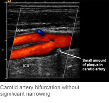
Carotid Arteries
Carotid ultrasound is used to evaluate the carotid and vertebral arteries in the neck for narrowing abnormal blood flow. This examination is painless and there is no radiation or intravenous dye.
Indications for Carotid ultrasound include:
- Symptoms of possible stroke or TIA
- Bruit
- Atherosclerotic disease elsewhere within the body
- Progression of previously known carotid narrowing
- Dizziness
- Hypertension
- Previous carotid artery surgery
How is carotid ultrasound performed?
While lying on a table, warm gel is placed on the neck, overlying the carotid arteries. Images are generated utilizing real time ultrasound which evaluates the blood flow in the carotid and vertebral arteries to determine if there is narrowing and blockage. Exam takes approximately 45-60 minutes.
Carotid Artery Screening
Carotid artery screening uses painless, non-invasive ultrasound imaging to image the carotid arteries in the neck for plaque and narrowing.
A stroke can result from the disruption of adequate blood flow to the brain. The most common source of a stroke is a significant narrowing or blockage of the carotid arteries caused by accumulation of fatty plaque along the carotid artery walls. The carotid arteries are the main blood supply to the brain, and plaque buildup in these arteries is the leading cause of stroke.
Indications for carotid artery screening:
- High blood pressure or currently taking medication for high blood pressure
- Currently a smoker or have a long history of smoking
- Irregular heartbeat
- High cholesterol or currently taking medication for high cholesterol
- Immediate family history of stroke or heart disease (mother, father, siblings, children)
- Exercise less than three times per week for 20-30 minutes at a time
- Diet high in saturated and/or animal fat
- Over 55 years of age
- Male
How is carotid artery ultrasound performed?
Gel is placed on the neck overlying carotid arteries. Images are generated utilizing real time ultrasound to determine if there is narrowing and blockage of the carotid artery. Exam takes approximately 30 minutes.
Venous ultrasound for thrombosis (DVT)
A lower venous duplex sonogram ultrasound study is performed using sound waves to evaluate the lower extremity for blood clots in the leg veins. This examination is painless and there is no radiation or intravenous dye.
Indications for DVT ultrasound include:
- Pain
- Swelling
- Previous deep venous thrombosis
- Recent joint replacement surgery
How is venous ultrasound performed?
Ultrasound gel is placed on the legs. Images are generated utilizing Doppler and color Doppler ultrasound, which evaluates blood flow. The test takes approximately 45 minutes.